 Plicae are some of the normal structures of the knee joint cavity Dupont JY. Synovial plicae of the knee. Clin Sports Med. 1997;16(1):87-122. They are folds of the capsule of the joint that surround the patella
(kneecap). The pleats are variable in size and structure between individuals. There
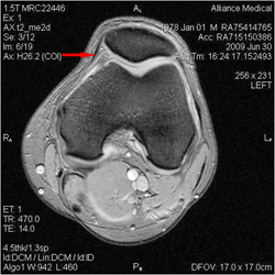
are at least 4 in most knees. The plicae are the "meniscus" of the patellofemoral joint. In this knee MRI the medial plica is indicated by an arrow on the inside of the left knee.
Knee Arthritis

Important educational information for Mr Hardy's patients on knee osteoarthritis or knee arthritis.
Click on the icons of patient information sheets for a searchable .pdf file to keep or print. Mr Hardy would prefer it if you saved the file to your computer for later reference than used paper resources.
When making an appointment in London or Bristol telephone Sally: 0044 (0)117 3171793.
Theatre Protocols for Knee Replacemen can be found below...
Theatre Protocols for Unicompartmental Knee Replacement, Patellofemoral joint Replacement can be found below...
Glossary
- Plicae
- Hoffa@s Fat Pad
- Arthritis
 The anatomy of the fat pad has been well described anatomically Gallagher J, Tierney P, Murray P, O'Brien M and radiologically Saddik D, McNally EG.
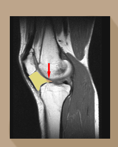
The red arrow points to the tongue of the fat pad (yellow) that catches between the condyle of the femur and the tibia. The condition, from chronic impingement then scarring of the fat pad, was first reported by German surgeon Albert Hoffa in 1904 (Albert Hoffa, 1859-1907) so it is odd that it has taken so long to recognise it as a common and genuine condition in athletes. Emad Y and Ragab Y.
The Hoffa's fat pad is known to be richly innervated with sensory fibres Wojtys EM, Beaman DN, Glover RA, Janda D. Innervation of the human knee joint by substance-P fibers. Arthroscopy 1990;6(4):254-63.
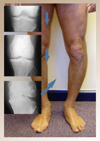
Arthritis is a blanket term for all causes of degenerative change in a joint. The most common cause world wide is mechanical impingement or trauma causing progressive degenerate change of the hyaline cartilage. The Image below shows the progression of radiographs from a normal knee that has suffered a neglected meniscal tear, then narrowing from degenerate change to frank osteoarthritis.
The definition of osteoarthritis is radiological and includes the features of loss of joint space, subchondral sclerosis, subchondral cysts and marginal osteophytes. Be careful to accept a radiology report that says osteoarthritis as many inexperienced radiologists over call what is seen on x-ray.
Mr Hardy's standard is to review his patients with their special investigations and the radiology report to hand so that symptoms can be compared with the x-ray and MRI findings. Anything less leads to incorrect diagnoses.
Knee Problems
Osteoarthritis of the Knee
There are four independant joints in the knee. Each of these joints can undergo degenerative changes that progress to osteoarthritis.
Mr Hardy is going to describe these conditions below and in the accompanying leaflets.
Osteoarthritis is due to mechanical breakdown of the surface hyaline cartilage of a synovial joint. In a normal knee the articulating surfaces of hyaline cartilage are congruent. The meniscus and plicae allow a spreading of the forces of load over a larger surface area and with flexion and extension presenting differing radiuses of the femoral condyle to the tibial condyle the function of the meniscus is to spread the load and reduce surface pressure.
Loss of this function happens in two ways. In an unstable meniscal tear a fragment of meniscus flips into the joint presenting a small area of fibrocartilage to the articulating hyaline cartilage.
The smaller the fragment the higher the pressure profile presented to the articulating surfaces (pressure =force/area).
Cyclic loading onto this small area with the high pressures causes progressive degenerate change of the surface of hyaline cartilage. The other way is by removal of the meniscus. Total or subtotal menesectomy increases the pressure on the hyaline cartilage by reducing the area through which the load is distributed. We know through various studies how potent menesectomy is at producing osteoarthritis.
During the progression of degenerate change to osteoarthritis the loss of this biomechanical function allows the concentration of forces. Cyclic loading of a degenerate joint appears to cause microfracture of the bone of the subchondral plate.
The attempt of healing of these microfractures is what causes the appearance of sub-chondral sclerosis as additional bone is laid down to heal the microfracture. Where the bone microfracture is not healed and where synovial joint fluid is forced through the microfracture subchondral cysts form. With the progression of inflammation associated with degenerate change come increases in new bone and cartilage formation at the margin of the joint.
This new bone and cartilage is called an osteophyte. Osteophytes limit the range of motion. They are often tender, can catch and cause pain on synovial folds and can break off to cause loose bodies which are painful when they cause impingement between articulating surfaces. A recent radiograph will help decide on the rate of degenerate changes taking place.
 Click on this image to read more...
It is important that Mr Hardy's patients understand that the majority of ordinary people he sees for a second opinion do not have this condition but are often in the stages of degenerative change that can be improved or reversed once the cause is identified and treated.
Osteoarthritis of the knee is a clinical diagnosis best made by a Consultant in Orthopaedics and Trauma. Knee osteoarthritis is characterised by the signs of inflammation (pain, swelling, heat and loss of function), stiffness, loss of range of motion and deformity.
The diagnosis is confirmed with an x-ray examination of the knee. Osteoarthritis is defined by four radiological (x-ray) features of advanced degenerative change. If you were given a report of and x-ray on your knee that says " there is some medial joint space narrowing and early osteophytes typical of osteoarthritis" them make sure your doctor has seen the radiograph itself as the variability in the differences between what is described and the grade of degenerative changes present are enormous.
Mr Hardy believes this careful definition of osteoarthritis in an individual is important because it has been established that the greater the impared ability to walk, the greater the risk of earlier death.
Arthritis of the Patellofemoral Joint
Osteoarthritis of the patello femoral joint is not uncommon. It starts with intermittent anterior knee pain and this becomes continuous knee pain. Crepitus (grating) of the patello femoral joint and osteophytes on the tibial spines (spiking) is an indication of early wear. Symptoms of difficulty rising from a chair or going up and down stairs can indicated advanced degenerate change. This initiating event appear to be due to either aplica syndrome and scarring or tear of the ligamentum mucosum and impingement of Hoffa's fat pad.
Arthritis of the Medial Tibiofemoral Joint
Osteoarthritis of the medial tibiofemoral joint is common. Most tibiofemoral osteoarthritis is due to an unstable meniscal tear.
Two thirds of all meniscal tears are medial (inside) one third of all meniscal tears are lateral (outside). Two thirds of all OA starts in the medial compartment and one third in the lateral compartment. The pattern of wear in the medial compartment depends on the aetiology of the wear as seen in population studies like the one comparing the pattern of varus wear in Middle Eastern patients with Western patients. Some patients develop pain and wear at the front of the inside of the knee from a prominent Hoffa's Posterior Fat Pad Impingement. Some patients get pain at the back of the inside of the knee from an unstable meniscal tear causing wear then degenerate change and then osteoarthritis.
For Mr Hardy's team, who are spending more time with patients preventing osteoarthritis of the knee, this pattern of wear observed in the paper above correlates with their findings in both groups of patients.
Arthritis of the Lateral Tibiofemoral Joint
Lateral meniscal tears usuall start off horizontal and stable. These can progress with further tearing to unstable tears that cause osteoarthritis through impingement.
Arthritis of the Superior Tibiofibular Joint
Osteoarthritis of the superior tibiofemoral joint is a rare and often missed diagnosis.
Treatment for Arthritis of the Knee
Osteoarthritis of the knee is treated conservatively or surgically.
The treatment for unremitting pain from osteoarthritis with an Oxford score of 20 or less where 48 is best function is knee arthroplasty.
However, Mr Hardy always helps patients to improve their quality of life and activities of daily living to avoid total knee arthroplasty until the benefits outweigh the risks.
He uses the Oxford Knee Score to help his patients decide on timeing for unicompartmental or total knee arthroplasty.
The first score on the left is in English.
The Oxford Knee score on the right is in Arabic.
Total Knee Arthroplasty
When all measures to first prevent osteoarthritis and then treat the impingement problems associated with osteoarthritis have been undertaken Mr Hardy recommends knee arthroplasty.
There are many types of arthroplasty including unicompartmantal knee replacement, total knee replacement, patellofemoral knee replacement, cemented knee replacement, uncemented knee replacement.
Mr Hardy will explain which is best in your circumstances. He has experience in unicompartmental, low contact stress, low wear surfaces, minimally invasive and navigation in knee arthroplasty. His team will discuss risks and benefits of these different procedures before you choose which would suit your circumstances best. Click on the icon below to read more....
Rheumatoid and Non-specific Arthritis
When I have time I will add to this section.
Gout
Gout can affect any joint, bursa, tendon or ligament that has suffered injury. The knee is no exception. Click on the image to read on....


